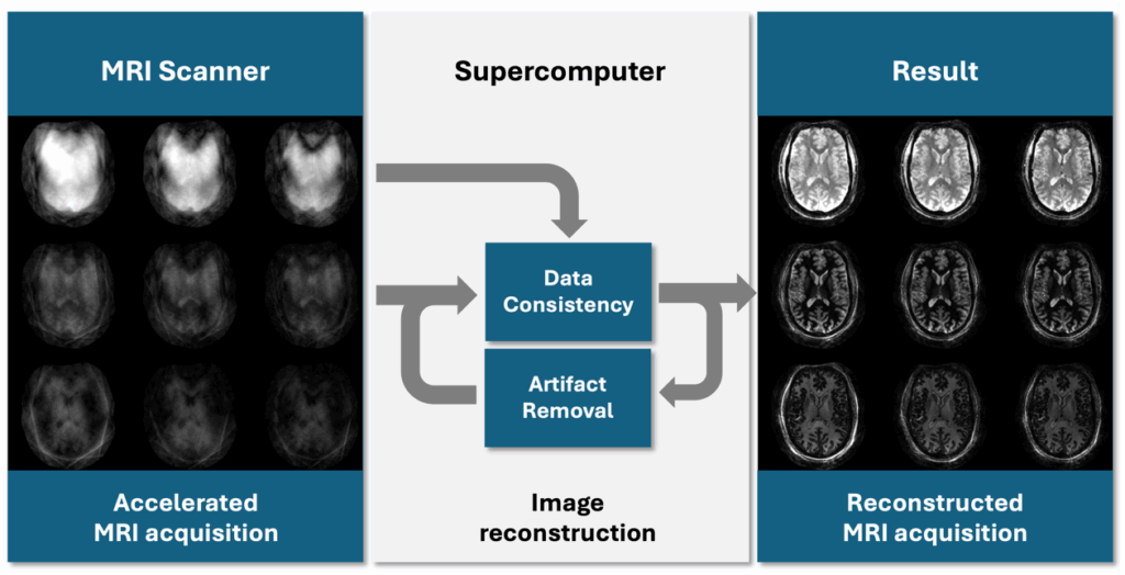
Technical/scientific Challenge
Quantitative MRI (qMRI) measures underlying MRI parameters, enhancing sensitivity to physiological changes and enabling reliable test-retest comparability, so that observed changes reflect true physiological differences rather than scanner variability. Translating qMRI to UHF, which produces higher-resolution imaging in shorter acquisition times, entails increased field inhomogeneities and specific absorption rate, though. Novel methods developed at INM-4 address these challenges but trigger significantly higher reconstruction complexity and prohibitively long reconstruction times.
Solution
To address these prohibitively long reconstruction times, INM-4, in collaboration with the Simulation and Data Lab Neuroscience, optimized its reconstruction code for HPC at JSC. By implementing efficient preprocessing, the reconstruction problem was converted into a slice-by-slice process that could be parallelized. Combined with automated slice processing via bash scripts, this reduced compute time from 320 hours to 8 hours per subject using HPC. This optimized workflow overcame previous limitations, enabling the application of a novel qMRI method that achieves faster scans, improved image quality, and precise parametric estimates. Subsequent measurement of a large cohort confirmed HPC-powered qMRI at UHF as a crucial step toward clinical feasibility.
Benefits
- HPC reduced reconstruction time from 320 to 8 hours per subject, making qMRI feasible.
- The optimised qMRI method enabled faster scans, improved image quality, and more precise parametric estimates.
- Collaboration with JSC enhanced computational efficiency and set the stage for AI-driven qMRI.
![Vergleichsdiagramm von Gehirn-MRT-Bildern, unterteilt in zwei Hauptabschnitte: Qualitatives MRT (links) und Quantitatives MRT (rechts). Unter „Qualitatives MRT“ gibt es eine Spalte mit drei graustufigen Gehirnabschnitten – axial (oben), sagittal (mittig) und koronal (unten) – die ein Strukturabbild mit willkürlicher Signalintensität zeigen. Eine Graustufen-Skala daneben reicht von -0,5 bis 0,5 und ist mit „Signal Intensity [a.u.]“ beschriftet. Unter dieser Spalte steht: „Structural image with arbitrary signal intensity.“ Der Bereich „Quantitatives MRT“ enthält vier Spalten, wobei jede die gleichen drei Gehirnansichten zeigt, jedoch mit unterschiedlichen Farbcodierungen und Messeinheiten: 1. Die erste Spalte zeigt farbkodierte Bilder von Blau (niedrig) bis Gelb (hoch), welche den „freien Wassergehalt in Prozent“ darstellen. Eine vertikale Skala rechts reicht von 0 bis 100 und ist mit „C_fw [%]“ beschriftet. Die Bildunterschrift lautet: „Free water content in percentage.“ 2. Die zweite Spalte zeigt Bilder in einer Farbskala von Blau/Grün (niedrig) bis Gelb (hoch) für die „Longitudinale Relaxationszeit in Millisekunden“, mit einer Skala von 0 bis 4000, beschriftet mit „T₁ [ms]“. Beschriftung: „Longitudinal relaxation time in milliseconds.“ 3. Die dritte Spalte verwendet eine Farbskala von Blau bis Gelb, um die „Effektive transversale Relaxationszeit in Millisekunden“ darzustellen, mit einer Skala von 0 bis 60, beschriftet mit „T₂* [ms]“. Die Beschriftung darunter lautet: „Effective transverse relaxation time in milliseconds.“ 4. Die vierte Spalte zeigt wieder Graustufenbilder, ähnlich wie beim qualitativen MRT, und stellt die „Magnetische Suszeptibilität in ppm“ dar. Die Skala reicht von -0,10 bis 0,10 und ist mit „χ [ppm]“ beschriftet. Darunter steht: „Magnetic susceptibility in ppm.“ Über den Bildern trennt eine waagerechte Linie die Überschriften: „Qualitatives MRT“ links und „Quantitatives MRT“ rechts. Jede Bilderspalte hat eine kurze, kursiv gedruckte Beschreibung des dargestellten Parameters und der jeweiligen physikalischen Einheit. Das Layout hebt den Kontrast zwischen einem einzelnen MRT-Scan mit willkürlicher Intensität und mehreren quantitativen MRT-Ansätzen hervor, die direkt kalibrierte, numerische Messwerte über die Eigenschaften des Gehirngewebes liefern.](https://supercomputing-in.de/wp-content/uploads/2025/08/QMRI-Fig2-1024x576.jpg)
Industrial sector
Health care / Pharmaceuticals / Medical devices, IT/HPC systems
Scientific partners involved

The Institute of Neuroscience and Medicine 4 (INM-4) at Forschungszentrum Jülich develops innovative methods to advance diagnostics and improve our understanding of the brain with state-of-the-art medical imaging technology, including ultra-high-field (UHF) 7T MRI. Its interdisciplinary approach in close collaboration with JSC leverages cutting-edge computational resources to develop novel imaging methods for visualizing new biomarkers at higher resolutions in shorter acquisition times.
Scientific impact
Implementing qMRI at UHF required a novel imaging method to address increased field inhomogeneities and specific absorption rate. This method enabled faster scans and improved image quality but required solving a complex reconstruction problem with high computational demands. Using conventional hardware, reconstruction was prohibitively slow—8 hours per slice for 160 slices per subject—delaying evaluation of its clinical potential.
To overcome this, the team turned to HPC and consulting services at JSC to adapt their software to harness HPC's power. Parallelizing tasks and automating processes reduced compute time from 320 to just 8 hours per subject, making it feasible to apply the novel qMRI method, which had been constrained by slow reconstruction.
HPC thus enabled the method’s practical use, improving image quality and parametric accuracy. This brings qMRI at UHF closer to clinical application, enhancing diagnostics. Further optimization—such as AI-driven image reconstruction—could eventually make it viable in routine clinical settings without direct HPC access, benefiting patients through more precise and timely diagnostics.
Read more
- „(ISMRM 2023) QRAGE – Multi-Echo MPnRAGE and Model-Based Reconstruction for Quantitative MRI of Water Content, T1, T2* and Magnetic Susceptibility at 7T“. Zugegriffen: 1. Oktober 2025. [Online]. Verfügbar unter: https://archive.ismrm.org/2023/1091.html
- M. Zimmermann u. a., „QRAGE—Simultaneous multiparametric quantitative MRI of water content, T1, T2*, and magnetic susceptibility at ultrahigh field strength“, Magnetic Resonance in Medicine, Bd. 93, Nr. 1, S. 228–244, Jan. 2025, doi: 10.1002/mrm.30272.